
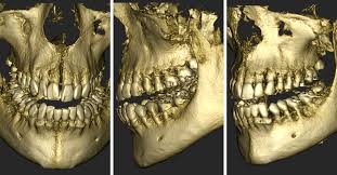
In modern dentistry, technological advancements continue to shape the way we diagnose, plan, and execute treatments. Among these innovations, Cone Beam Computed Tomography (CBCT) has emerged as a cornerstone technology, using 3D images to provide detailed insights into the intricate structures of the maxillofacial region.
Introducing a CBCT to your dental practice can have transformative benefits for treatment planning and your work in endodontics, orthodontics, or implantology. As a dental professional, the decision to integrate new technologies into your practice is pivotal, and you want to know you are making the right choice. So, what makes a CBCT so valuable?
Universal benefits
Investing in a CBCT imaging system in your dental practice will bring a multitude of overlapping benefits across endodontics, orthodontics, and implantology, including:
- Detailed visualisation
- Accurate assessment
- Precise treatment planning
- Enhanced patient communication
- Efficiency and workflow optimisation
By leveraging the capabilities of CBCT imaging, you can deliver personalised, evidence-based care that ensures optimal clinical outcomes and patient satisfaction, ultimately elevating the standard of your dental practice.
CBCT in endodontics
In endodontics, precision is paramount and complexities common, CBCT can be an invaluable tool. With its ability to provide high-resolution, 3D images of the root canal system and surrounding structures, CBCT offers insights that go well beyond what traditional 2D radiography can offer.
One of the most significant advantages of CBCT in endodontics lies in diagnostic accuracy. By offering detailed visualisation of root canal anatomy, periapical lesions, and tooth fractures, CBCT empowers endodontists to uncover hidden complexities and anatomical variations that may remain hidden from conventional radiographic techniques.
Furthermore, CBCT plays a pivotal role in treatment planning, enabling endodontists to develop tailored strategies. From identifying accessory canals and calcified root canals to assessing the proximity of adjacent anatomical structures, CBCT equips endodontists with the comprehensive information needed to navigate treatment complexities with confidence.
Moreover, CBCT imaging enhances procedural guidance during root canal therapy. By visualising the intricate anatomy of the root canal system in three dimensions, endodontists can navigate canals with greater accuracy, reduce the risk of procedural errors, and improve treatment outcomes.
CBCT in orthodontics
Precision is paramount in orthodontics. The intricate interplay of dental and skeletal structures demands meticulous planning and execution to achieve the best treatment outcomes. This is where CBCT can be a game-changer.
CBCT imaging provides orthodontists with detailed 3D views of the dentofacial complex, allowing for precise assessment of tooth position, root morphology, and skeletal relationships. This level of insight enables practitioners to diagnose orthodontic issues with accuracy, from impacted teeth and dental anomalies to skeletal discrepancies.
One of the key advantages of CBCT in orthodontics lies in its ability to facilitate treatment planning with a focus on individualised care. Whether it's determining the ideal bracket placement, predicting tooth movement, or planning for orthognathic surgery, CBCT empowers orthodontists to make informed decisions that align with each patient's goals and anatomical characteristics.
Moreover, CBCT imaging plays a pivotal role in assessing the feasibility and safety of orthodontic interventions, particularly in cases involving impacted teeth or complex skeletal discrepancies. By visualising the spatial relationship between teeth, roots, and adjacent structures, orthodontists can anticipate potential challenges and devise strategies to mitigate risks before initiating treatment.
Beyond diagnosis and treatment planning, CBCT enhances patient communication and engagement. Patients can better understand the rationale behind their treatment plan and see the anticipated outcomes. This fosters trust and confidence in the orthodontic treatment journey, leading to greater patient satisfaction and compliance.
CBCT in implantology
With CBCT imaging providing detailed 3D views of the bone, nerves, sinuses, and adjacent teeth, implantologists can assess bone quality, quantity, and morphology with exceptional accuracy, guiding the decision-making process regarding implant placement and ensuring the optimal functional and aesthetic outcomes.
In implantology, a CBCT can facilitate virtual implant planning. Using specialised software, dental professionals can simulate the placement of dental implants on the computer-generated 3D model of the patient's jawbone. This virtual implant planning enables precise determination of implant position, angle, and depth, taking into account anatomical considerations and prosthetic requirements. By meticulously planning implant placement prior to surgery, CBCT minimises the risk of complications and enhanced the predictability of treatment outcomes in implantology.
CBCT imaging also plays a crucial role in assessing the feasibility of implant placement and identifying potential anatomical constraints such as inadequate bone volume or proximity to vital structures. With a precise 3D image, implantologists can anticipate potential challenges and devise strategies to address them effectively, whether through bone augmentation techniques or alternative implant placement approaches.
Beyond the surgical phase, CBCT imaging facilitates the fabrication of custom surgical guides. Additionally, CBCT enables postoperative assessment of implant integration and peri-implant bone health, allowing for timely intervention if complications arise.
Which CBCT is best for your practice?
Buying a CBCT is a large investment, and knowing which one is best for your unique dental practice can be difficult. Read about two versatile and advanced models, the NewTom GO and the Acteon X-MIND Prime 2, and see what they can do for your dental practice.
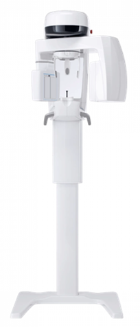
NewTom GO
The NewTom GO offers high-resolution images, exceptional performance, and safety features such as low-dose protocols and SafeBeam™ technology.
- Endodontics: Its advanced imaging technology and low-dose protocols enable precise diagnosis of root canal anatomy and periapical lesions.
- Orthodontics: With superior image quality and stable patient positioning, the NewTom GO supports accurate assessment of dental and skeletal structures.
- Implantology: The NewTom GO's Hi-Res mode and autoadaptive Metal Artifact Reduction (aMAR) algorithm ensure clear visualisation of anatomical structures.
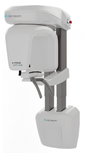 Acteon X-MIND Prime 2
Experience the power of simplicity in dental imaging with the Acteon X-MIND Prime 2, a multi-function imaging system combining 2D panoramic, cephalometric, and CBCT imaging for optimal dental procedures.
- Endodontics: High-resolution imaging and the Selective Metal Artifacts Reduction Tool (SMAR-T) ensure precise diagnosis of root canal anatomy and periapical lesions.
- Orthodontics: The X-MIND Prime 2's comprehensive exam modalities and excellent image quality enable accurate assessment of dental and skeletal structures for treatment planning and evaluation.
- Implantology: Simplified surgery planning with the Implant Planning Wizard and Cloud implant library, combined with high-resolution imaging and advanced detection technologies, ensures accurate assessment of bone quality and quantity for dental implant placement.