Digital X-ray Imaging
Digital X-ray imaging systems take accurate X-rays of patients’ oral anatomy in 2D or 3D. Accurate and detailed 3D images are now achievable at ever lower doses making it possible to ensure accurate diagnosis and patient safety simultaneously.
Introduction
X-rays have always been a vital part of diagnosis and treatment planning in dentistry. For many treatments, such as implant dentistry and endodontics, an accurate and clear view of the oral situation beneath the teeth and gums is essential to ensure optimum, safe treatment outcomes.
This interactive guide takes a deeper look into dental digital X-ray imaging and the different options available. What have been the recent developments in digital X-ray imaging and what are its applications in modern dentistry? What should you consider when purchasing a digital X-ray machine?
What is digital X-ray imaging?
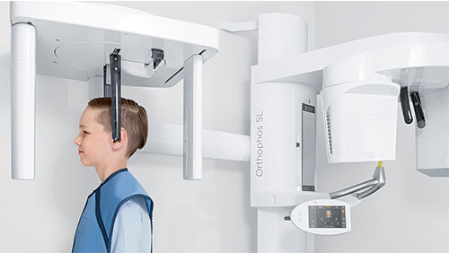
X-ray imaging is the taking of X-rays of the mouth to generate images of the tooth and bone structure, soft tissue and nerve paths underneath the gums. Digital X-rays are taken by machines which have a digital image sensor which captures the X-ray and displays them on a digital screen. They are fast replacing traditional film X-rays due to the lower doses of radiation required and the impressive level of detail which can be achieved with digital technology.
There are two types of digital X-rays commonly used in dentistry, 2D digital X-rays and 3D X-rays. Traditionally in an effort to keep radiation doses as low as reasonably achievable and because 3D X-rays have until now required much larger doses of radiation, 2D digital X-rays have been widely used.
However, the technology behind Cone Beam Computed Tomography (CBCT) scanners, which are used to take 3D digital X-rays, has improved markedly recently and it is now possible to take extremely clear and detailed images at very low doses of radiation.
The benefits of 3D digital X-rays
-
A 3D view
3D is often preferable to 2D as it shows depth as well as height and width. With 2D there is always an element of guesswork as the exact location of structures cannot be determined. Whilst 2D images are perfectly adequate for evaluating tooth decay, bone loss and other more common dental issues, 3D is essential for more complicated procedures such as implant placement and endodontics.
-
Outstandingly clear images
It is possible to achieve extremely clear, high-definition images with a digital 3D imaging machine which gives the clinician all the information they need to effectively diagnose and plan treatment. The technology behind digital X-ray machines has advanced significantly to enable the capture of sharp, accurate representations of patients’ oral anatomy in both 2D and 3D.
-
3D at low dose
The real game-changer in recent developments in 3D digital X-ray technology is the ability to capture 3D images at extremely low radiation doses. This has removed a common barrier for many clinicians who have discounted 3D images due to the higher radiation doses required.
Dental practices are required to adhere to both the Ionising Radiation (Medical Exposure) 2000 Regulations (IR(ME)R) and the Ionising Radiations Regulations 2017 (IRR17) , which are based on the ALARA principle of only exposing a patient to a radiation dose that is “as low as reasonably achievable.” As a rule, dental practitioners should favour a lower dose wherever practicable, as long as the resultant X-ray provides the detail and information required to make an accurate diagnosis and an effective treatment plan. -
Better diagnosis
An accurate picture of the oral situation is key for diagnosis, especially for more complex treatment such as endodontics and implant surgery. For instance, it enables endodontists to determine much more easily the presence and location of disease and therefore decide upon the long-term prognosis of the tooth.
-
Better treatment planning
A clear 3D model of the oral anatomy enables clinicians to plan treatment much more effectively. Implant surgeons can determine the optimum placement and angle of the implant, taking into account bone density and the location of any nerves. Endodontists can accurately plan their access and ensure they know the number and configuration of root canals and the curvature of the roots. 3D removes the element of surprise.
-
Efficiency
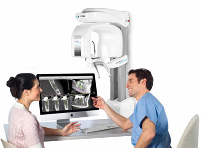
From accurate diagnosis and comprehensive treatment planning comes efficient treatment. The clinician is ready with the requisite tools and materials to carry out a planned and optimised treatment and this leads to better patient outcomes. Having a CBCT scanner in-practice with both 2D and 3D capabilities, and a range of dosage and field of view options means the clinician is fully equipped to make the right treatment decisions in the shortest possible time frame - which benefits the patient and the business.
Applications for digital X-rays
-
Implant surgery
Crystal-clear 3D X-rays are essential for implant placement planning. Only then can the clinician be sure they are placing the implant in the optimum position in terms of bone density and implant angulation, whilst avoiding any nerves or other anatomy.
A detailed 3D view also gives the implant clinician an accurate picture of any pathology and possible complications that might arise from it. An area for possible concern on a 2D image might look relatively small, but in 3D it may appear more significant. The exact location of any nerves will also be clearer.
-
Orthodontics
Extraoral X-rays reveal the relationship of the teeth to the jaw and rest of the head. They help orthodontists identify impacted teeth, monitor growth and development of the jaws and to identify potential issues between teeth, jaws and the temporomandibular joint.
A 2D orthopantogram (OPG) is commonly used in orthodontics to obtain a panoramic view of all the teeth in the lower and upper jaws in a single film, thereby demonstrating the number, position and growth of teeth, including teeth that have not surfaced yet.
Cephalometric X-rays show the entire side of the head, useful for examining the teeth in relation to the jaw and profile of the head.
CBCT X-rays provides the orthodontist with unrivalled accuracy particularly in relation to the three-dimensional location of different parts of the oral anatomy. It provides accurate measurements in 3D, improving such things as the localisation of impacted teeth, the existence of any airway abnormalities and the exact measurement of any asymmetry.
-
Endodontics
3D digital X-rays in particular help the endodontist to accurately assess the clinical situation and diagnose any disease. This facilitates their decision on the best treatment and then helps them plan the treatment meticulously to ensure that no root anatomy is missed and that the best access points are identified. It takes the guesswork out of endodontic treatment planning and ensures the clinician is primed and ready for the treatment with the best tools at their side.
How to evaluate digital X-ray machines
There are many factors to consider before investing in a digital X-ray machine. All of the following are recommended criteria:
√ Open and therefore able to be used by a variety of dental equipment and software
√ Clinically proven
√ Cost-effective
√ No ongoing fees or licences
√ Long warranty
√ Future-proof / easily upgradeable
√ Offers a range of definition settings and low dose options to cover all applications
√ Offers a range of fields of view to ensure adherence to the ALARA principle
√ Easy to use, including straightforward patient positioning
Browse our two best-selling digital x-ray machines

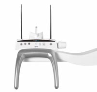
Acteon X-MIND® Prime