
The power of digital scanning and X-ray imaging combined
Digital impressions and digital X-rays both deliver extraordinary accuracy and clarity and have revolutionised impression and X-ray taking respectively. But what happens when the two are combined?
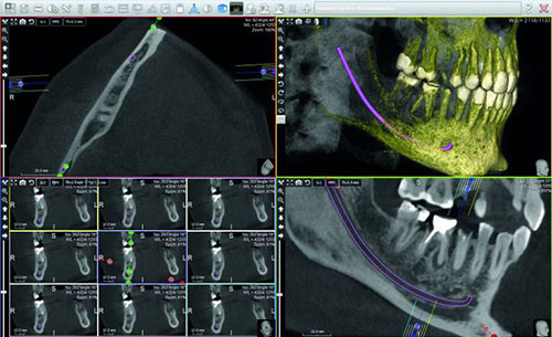
CAD/CAM software can merge the surface scan from a digital impression with a 3D digital X-ray to give the clinician a complete picture of the oral situation above and below the gum line. This is invaluable for some areas of dentistry in particular and also for communication both with the patient and within the dental team.
Implant-borne restorations
Combining a surface impression from an intraoral scanner and a 3D digital scan together gives an implant surgeon all the information they require to plan the placement of one or more implants and a restoration on top. The surface impression and digital scan are imported into CAD/CAM software and merged. The restorative clinician designs the restoration within the software and plans the implant placement concurrently ensuring they work seamlessly together. They can even design a surgical guide within the software and print it out on a 3D printer, or order it along with the restoration from the laboratory. This means that implants can be placed and the prosthetic manufactured with pin-point accuracy which results in an optimally fitted, aesthetic and long-lasting restoration.
Orthodontics
Digital impressions and 3D digital X-rays can be used together to deliver custom, patient-specific orthodontic appliances - even in-house with the addition of a 3D printer. A 3D radiographic view of the patient’s anatomy means the clinician no longer has to guess the 3D location of an impacted tooth or the correct eruption vector. Merging the X-ray and the surface scan enables the clinician to use the root anatomy to devise the correct orthodontic treatment plan, improving the long-term outcome.
Patient communication
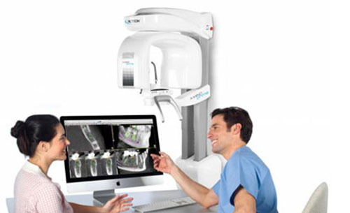
The ability to show the patient images of their oral anatomy both above and below the gums gives patients a much better understanding of the diagnosis and the intended treatment plan. Accurate pictures speak more than a thousand words which significantly improves patient understanding and therefore treatment uptake.
Dental team communication
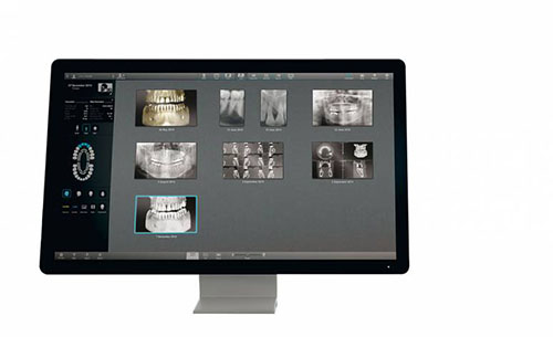
Digital images and CAD/CAM software have succeeded in improving communication between the whole dental team, including clinician, technician and any third-party partners. Communication is instant and misunderstandings are avoided because every team member has immediate access to the detailed images and design files. From a patient record-keeping point of view, digital images make it much easier to keep all patient information together in a single file on a practice management system for instance. Diagnostic images can then also be tracked more easily to monitor disease progression or any other changes in the patient’s dentition.