Magnification

The use of magnification in dentistry to improve the visual acuity of the oral cavity is essential for early diagnosis and more accurate treatment. Magnification devices such as surgical microscopes, loupes and lights enhance vision and improve ergonomics and can be used in many areas of general dental practice including:

  • restorative dentistry
  • endodontics
  • fixed and removable prosthodontics
  • periodontics
  • oral surgery

This interactive guide takes a deeper look into loupes, microscopes and the different options available. What are the advantages and what are their applications? What should you consider when purchasing loupes or microscopes?

Magnified image

Magnified image

Brilliant illumination

Brilliant illumination

Better posture

Better posture

Improved comfort

Improved comfort

Increased precision

Increased precision

Improved patient care

Improved patient care

Improved efficiency

Improved efficiency

Loupes & Lights

Loupes are the most commonly used devices for magnification in dentistry, improving direct and indirect vision for clinicians, hygienists and therapists without major changes to working protocols. Loupes are available in a range of styles and magnifications, the most common magnification being 2.5x, providing an ideal balance between visual acuity and field of view.

FIND OUT MORE

Microscopes

Dental operating microscopes are widely used in dentistry to provide magnification and illumination for clinicians to see fine details within the oral cavity not visible to the naked eye. Microscopes allow for more accurate treatment planning and execution while providing better ergonomics for clinicians to reduce the risk of musculoskeletal disorders.

FIND OUT MORE

Introducing Loupes

Magnification was introduced into dentistry as far back as 1876 and loupes are now the most common way of magnifying the treatment area. Growing in popularity, along with the increase in minimally invasive techniques, loupes enable clinicians to not only see in more detail, which has clear benefits for diagnosis and treatment, but also lets the clinician work at the optimal distance from the patient with the resultant benefits for posture and ergonomics.

loupes intro

Why use loupes?

Regardless of the type of treatment offered, loupes are an essential piece of equipment for all clinicians. No longer just the preserve of specialists, in endodontics and minimally invasive dentistry, many dentists routinely wear loupes for every patient and procedure.

VIEW ALL HENRY SCHEIN LOUPES

The Benefits

The technology has advanced significantly since the beginning and dental loupes now offer clinicians the following compelling benefits:

Clarity of vision:

The extra clarity gained from wearing loupes and using a suitable light makes accuracy and attention to detail easier to achieve.

Diagnosis:

Being able to see the treatment area in detail assists with accurate diagnosis, by being able to see the treatment area in close detail.

Ergonomics:

It is well known that when carrying out dental procedures, poor posture can contribute to back and neck pain. Many clinicians suffer from chronic back pain and there is evidence that the use of appropriate magnification can help resolve some of these issues.

Posture:

Achieving the correct operating posture can be helped by the wearing of loupes as the magnification allows the operator to maintain the optimal working distance from the patient.

REQUEST MORE INFO

Which loupes are right for me?

Choice of loupes is very much a personal matter and there is a whole range of different designs, frames and specifications to choose from. Here are some of the things you should know before choosing your loupes.

Depth of field

This refers to the ability of a lens to focus on objects, both near and far, without needing to change position. When magnification is used it restricts the depth of field and as the magnification increases the depth of field decreases, until a point at which only a small object is in sharp focus. At high magnification just a small movement from the patient or the clinician will result in losing focus, making working more challenging.

High-performance oculars can provide an extra-large depth of field (up to 120 mm).
• Short 28-38cm
• Regular 36-46cm
• Long 43-53cm
• X-Long 50-60cm

Depth of field

Optical quality

When purchasing loupes you should also consider the following…

Resolution:
The ability to differentiate images that are separated by small distances and to detect fine details.

Colourmetric Shift:
Longitudinal Chromatic Aberration, also known as “LoCA” or “bokeh fringing”, occurs when different wavelengths of colour do not converge at the same point after passing through a lens. Because each colour has a different wavelength, uncorrected optics cannot focus on the different wavelengths in different points in space.

Deformation of the image:
An object that is seen with a magnifying glass that has a spherical aberration, appears bent or spheric. The lines are not straight.

Field of view

The basic principle for field of view is that as the magnification increases, the field of view decreases. In practical terms a magnification of 2x – 2.5x would enable the clinician to see multiple quadrant areas in focus and this is the magnification used by most general practitioners. Once a magnification of 3.5x is reached, the field of view becomes more restricted, usually to a single quadrant. However, this ability to focus on a particular area, often on a single tooth, makes this type of magnification suitable for specific procedures such as endodontics.

Field of view

Weight

Weight and balance are vital for operator comfort and this is an important consideration for those wearing loupes constantly throughout the working day. The type of lens (Galilean or Prismatic) and design of the loupes all play a part in weight and balance considerations.

Lenses

Galilean Lens:
This is the most common type offering magnification of 3.5x or less. The lightweight design offers good field-of-view and good depth-of-field in a short compact design.

Prismatic Lens:
Much higher magnification (up to 8x) provides the sharpest image possible, within a short depth-of-field. However, this is compensated by longer and therefore heavier lenses. These are suitable for specialists requiring great detail and clarity within a small area.

Range of Loupes

PeriOptix® Through-the-Lens Loupes

PeriOptix optics are made with high-end, shatter-resistant glass to ensure optimum edge-to-edge clarity in a large viewing field. These lightweight premium through-the-lens (TTL) loupes have extremely precise optical quality and a large field of view making them comfortable, easy to use, and carefree.

  • Adjustable to all users
  • Variable declination angle
  • Less expensive (-15 to 20%)
FIND OUT MORE
REQUEST MORE INFO
PeriOptix® Through-the-Lens Loupes

Frames

Classic Titanium Frames

No feature is more important for comfort of the operator than the frame. PeriOptix frames have a wide variety of styles and adjustments, providing comfort, customisability and style.

REQUEST MORE INFO

Let there be light

An appropriate light source is essential if true image clarity is to be optimised. PeriOptix improves visual acuity through powerful headlights that remove shadows in the oral cavity and eliminate the need to constantly adjust overhead lighting. Good lighting provides uniform and natural-colour output that is crucial to ensure the best outcome for your patients.

Firefly® Cordless Headlight System

Easily upgrade your loupes to incorporate a Firefly cordless headlight and enjoy the freedom to move through any procedure with confidence.
  • Eliminates heavy power packs and annoying cords
  • Easy to clean, reducing the risk of cross-contamination
  • Provides a well-defined and uniform spot with natural colour output
  • Light Intensity: 2 settings — high 32,000 lux and low 20,000 lux
  • Weight: 28 grams
  • Field Illumination: 70 mm
  • Available in black, red and blue
FIND OUT MORE
REQUEST MORE INFO
Firefly® Cordless Headlight System

LumiPro Headlight System

This innovative light (with cord) maximises clarity, ergonomics and control alongside the following benefits:

  • Uniform output of natural, bright light
  • Enhanced spot size to illuminate the entire oral cavity 
  • Significantly smaller and lighter than most headlights in the market
  • Easy-to-use soft-touch on/off and intensity adjustments to reduce the need to deglove
  • 10+ hours of run time on high
FIND OUT MORE
REQUEST MORE INFO
LumiPro Headlight System

Measurements

Being measured accurately for your loupes is vital and two measurements are normally taken.

REQUEST MORE INFO

Pupillary Distance (PD)

This is the measurement, in millimeters, between the centers of the pupils in each eye. This measurement only takes a couple of minutes.

Pupillary distance

Working Distance

This is the measurement which determines the distance at which the user is comfortable working. This distance should be calculated while the user is in an upright, and comfortable position that keeps the clinician’s ergonomics in check.

Working Distance

Introducing Microscopes

Microscopes are particularly useful in dentistry for various purposes such as diagnosis of subgingival deposits, microfractures, longitudinal fractures, defective marginal fit of restorations and other things that can be overlooked clinically. In addition, magnification is often necessary to study root canal anatomy and the root surface in endodontics.

In general magnification can be set to between 3x and 30x, thereby expanding the diagnostic options due to better lighting and sight.

When coupled with the appropriate capture devices, microscopes can become instruments capable of projecting and recording clinical procedure in a more streamlined and efficient manner.

Dr Milen Dimitrov

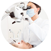
There are five key areas in which microscopes offer distinct advantages and benefits to the clinician:

  • Magnification
  • Illumination
  • Precision
  • Visualisation
  • Ergonomics

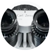
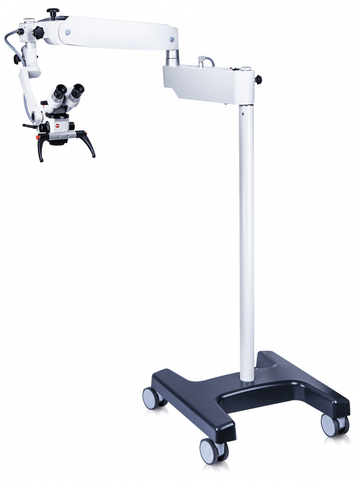
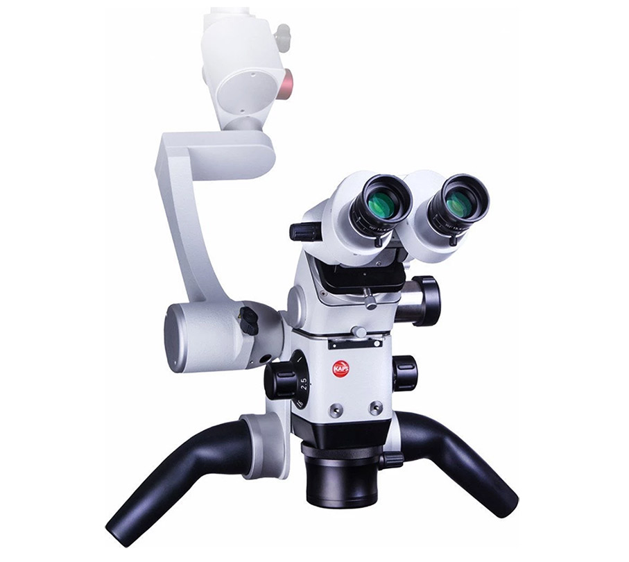
Magnification: this is determined by the power of the eyepiece, the focal length of the oculars, the magnification changer and finally the focal length of the lens.

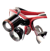
Microscopes normally provide between 3x and 30x magnification with the ability to change views. Microscopes offer a deeper depth of field of view and a wider field of view than dental loupes.

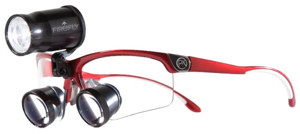
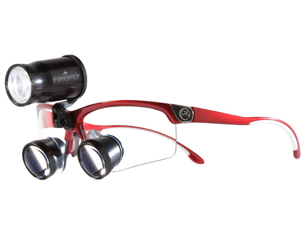
Illumination: When magnification is used, the available light is spread out and the image appears darker. To compensate for this, an LED light, mounted directly on the microscope head provides a bright, white light.

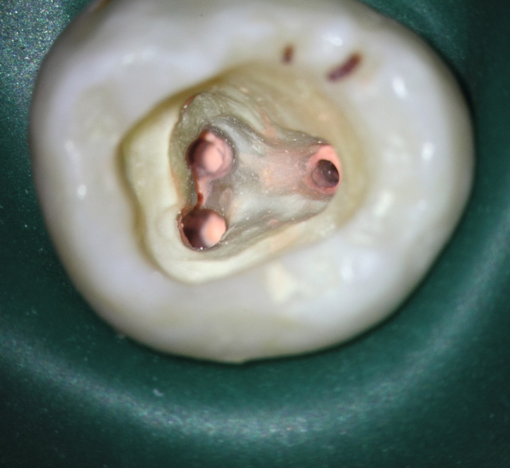
Precision: Increased targeted magnification using a microscope enables dentists to see the treatment area in great clarity and therefore aids the completion of detailed work.

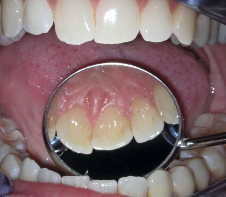
Visualisation: Dentists can only treat what they see. When using a microscope, the lens can be positioned directly over the patient’s mouth without any additional discomfort either for patient or clinician.

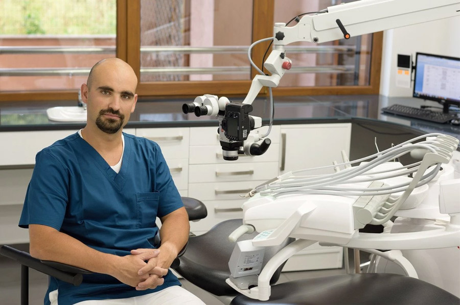
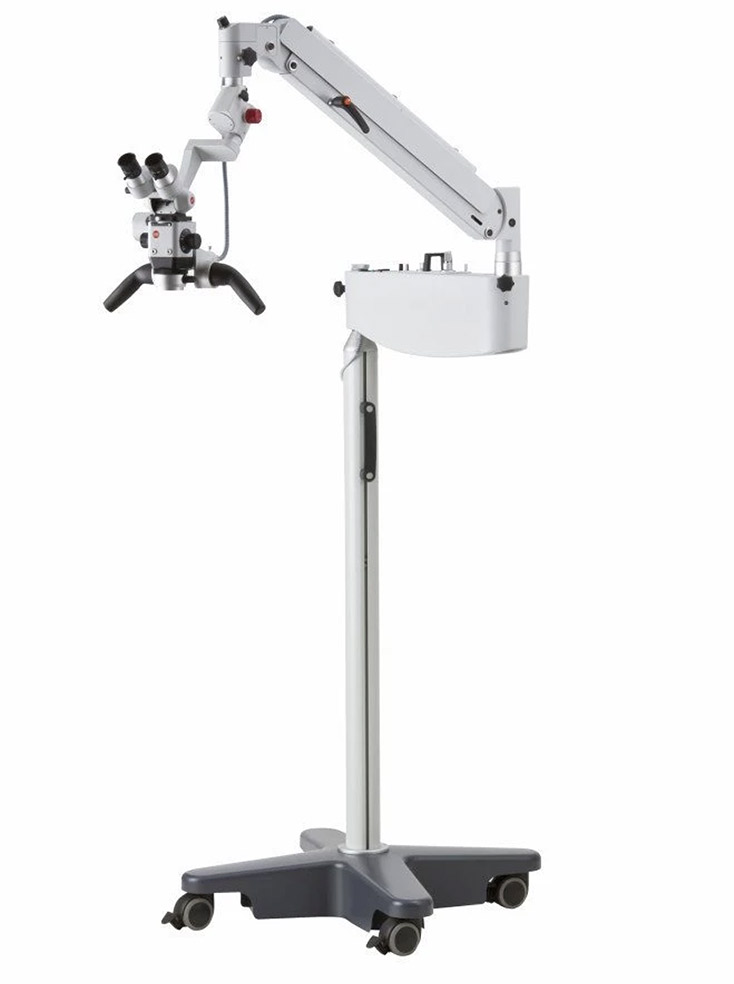
Ergonomics: When using a microscope dentists can be seated always upright in an ergonomically healthy position, which means a more comfortable working environment, particularly for longer procedures. This is aided by microscope heads that swivel laterally as well as moving horizontally. Some treatment centres also have slow motion movement which helps this even more.

Microscope Applications

The benefits of microscopes are many and varied and have applications across a range of dental disciplines.

VIEW HENRY SCHEIN DENTAL MICROSCOPES

Restorative Dentistry:

  • Provides refinement in tooth and margin preparation
  • Allows for closer inspection of restorations and marginal tissues
  • Helps with inspection of marginal fit of the restoration
  • Improved lighting and magnification help with caries detection and removal
  • Improves detection and evaluation of coronal and root fractures and other abnormalities
  • Facilitates cord placement for gingival retraction
  • Enables better inspection of impressions
  • Assists in gingival contouring or reshaping around teeth and implants

Endodontics:

  • Improves visualisation of fracture patterns, both of the coronal aspect of the tooth, the chamber, and into the root structure.
  • Enables the isolation, evaluation, and remediation of pulpal chamber obstructions, calcifications, and resorptions
  • Enables predictable pulp chamber visualisation
  • Three-dimensional visualisation of root obstruction from separated instruments allowing for the successful removal of blockages
  • Enables the thorough cleaning of deep caries, which can be completely illuminated, removed, and sealed
  • Easier manipulation of gingival tissue
  • Reduces operator fatigue and eyestrain during procedures

Periodontics:

  • Clinician can minimise the size of the surgical site, reducing patient discomfort and healing time
  • Improves accuracy of microsurgical incisions and suturing
  • Allows for better inspection and diagnosis of abnormal soft tissue lesions
  • Aids identification of micro-inflammation following non-surgical therapy
  • Helps inspection of the quality of restorations and marginal tissues
  • Improves visualisation of implant sites and helps in evaluating the exact fit of prosthetic components
  • Permits precise control of laser surgery
  • Facilitates sinus lift procedures through direct visualisation of the sinus membrane during dissection
  • Provides upright working conditions, alleviating neck, back and shoulder problems

Prosthodontics:

  • Easy diagnosis of microscopic cracks in teeth
  • Enables micro adjustments of crowns and bridges during ‘try-in’
  • Useful in the finishing and polishing of the final edge of prostheses
  • Aids the achievement of high quality aesthetic outcomes
  • Enhanced visualisation simplifies complicated treatments

Surgical:

  • Powerful magnification can be used to examine very fine details for diagnostics and intrasurgical examinations
  • Postural, procedural, psychological and educational benefits for clinicians and patients
  • Enables clear vision to aid all surgical treatments, including implants, impacted teeth and soft tissue grafting
  • Of particular value in the surgical treatment of injuries and lesions in the sensitive area of the mouth, including lingual nerve and lower jaw nerve
  • Procedures using microscopes involve less operative trauma and therefore improved healing and outcomes

Which Microscope is right for me?

Kaps 900

The Kaps 900 is a high performance and user-friendly dental microscope. It supports the dentist’s routine work and is a perfect entry level model with an attractive price-performance ratio and the option of having the microscope mobile, wall mounted or ceiling mounted (ceiling permitting).

Brilliant Optics

Kaps optics deliver crystal-clear images and high resolution details in natural colours (apochromatic optics).

Integrated LED Lighting

The high-contrast colour temperature of the LED lighting strongly resembles daylight. In addition, the even lighting of the field of vision makes work less tiring. Further advantages include the long durability (50,000 hours) and low electricity consumption. The LED lighting is noiseless and generates minimum heat.

Ergonomics

With the Kaps 900, clinicians are always seated comfortably in an ergonomic upright position, resulting in a much more relaxed approach to clinical work.

  • 5-step magnification changer
  • 0-210° inclinable binocular tube f = 182 mm
  • Objective f = 250 mm, with fine focussing
  • Eyepieces 12.5 x V
  • Integrated LED-illumination, dimmable (operating time circa. 50,000 hours)
  • Space-saving mounting systems
  • Ergonomic hand grips

Technical data

  • Magnification – 5-step apochromatic magnification changer
  • Binocular tube – 0-210° inclinable binocular tube f = 182 mm
  • Eyepiece – Wide field eyepiece 12.5 x V; 10 x V
  • Objective
  • Objective f = 250 mm with fine focusing
  • Kaps-Variflex-100 f = 220 mm – 320 mm (continuously)
  • Integrated LED illumination
DOWNLOAD THE BROCHURE
Microscope Kaps 900
Microscope Kaps 900

Kaps 1100

The Kaps 1100 is an advanced, powerful and user-friendly dental microscope with lateral head movement, a magnification unit with 5 step changer, optical variflex lens and a monolateral beam splitter. This gives the ideal combination of solid build, reliable usability and outstanding optics. In addition, the lateral head movement allows for optimal seating ergonomics.

Microscope Kaps 1100 variflex

Brilliant optics

The Kaps 1100 delivers crystal-clear images and high resolution details in natural colours (apochromatic optics). Whether progressively with a changer system or continuous with a zoom system, the Kaps 1100 provides a general overview at low magnification as well as clear detail recognition at higher magnification.

Ultra-bright LED illumination

The LED lighting enables a view into areas normally difficult to access. The high illumination, significantly better contrast and colour temperature in daylight quality reduces work fatigue. The lighting system is highly energy-efficient while the long lifetime of the LEDs guarantees lower operating costs.

Balancing system

Using the balancing system, the Kaps 1100 can be adjusted smoothly and precisely to any position, even when adapted accessories are used.

Ergonomics

Working with the Kaps 1100 is always done in a comfortable, upright and ergonomically correct position, preventing back and neck pain even with long treatment times.

The perfect all-rounder

  • 5-step apochromatic magnification changer
  • Binocular swivel tube 0 – 120
  • Objective f = 250 , with fine focusing (12 mm)
  • Eyepieces 12.5 x V
  • Balancing system
  • Ergonomic handles
  • Integrated LED illumination

Technical data

  • Magnification: 5 step apochromatic magnification changer
  • Modification range: 1.5x – 40x available (dependent on objectives and eyepieces)
  • Field of view: 120 mm – 7 mm (dependent on objectives and eyepieces)
  • Illumination: Integrated LED illumination
    • Optional: Xenon 180 W (external)
    • Integrated variable illumination spot size and orange filter
  • Objectives:
    • Fixed focal length from 100 mm to 400 mm offered in 50 mm increments, each with fine focusing 12mm
    • Kaps-Variflex-100 f=220 mm – 320 mm (stepless)
    • Vario Objective (Modul 50) f=200 mm – 400 mm (stepless and motorised)
  • Eyepieces: 10 x; 12.5 x
FIND OUT MORE
Microscope - Kaps 1100

Dental microscope light improves visibility during light-curing composite application white paper:

DOWNLOAD NOW

Clarity of vision is vital to meet the demands of today’s patients. Loupes, lights and microscopes are an important part of a clinician’s equipment and create the right environment to deliver outstanding treatment. There has never been a better time to invest in magnification and Henry Schein Dental has picked the best equipment to help clinicians make the most appropriate choice.

Talk to a Henry Schein representative now

REQUEST MORE INFO

Invest in your future

The power of magnification to transform dentistry cannot be disputed. The high quality of treatment that is made possible by loupes and microscopes is now well-documented and there is no doubt that they will play a significant role in the future of dentistry.

Accurate diagnosis and confident treatment planning are the foundation of successful treatment outcomes and the latest equipment now makes both consistently achievable. There has never been a better time to invest and Henry Schein Dental has picked the best equipment to help clinicians maximise their potential.

Our Capital Asset Finance schemes can help you to maximise the Annual Investment Allowance, where you can offset up to £1,000,000 of capital asset spend against pre-tax profits in any full financial year. In addition to a variety of finance options, we also offer very competitive Flexlease plans on our larger equipment.

TALK TO A HENRY SCHEIN REPRESENTATIVE NOW